Home

balsa agrio Teórico raman imaging Posible Ru En la mayoría de los casos

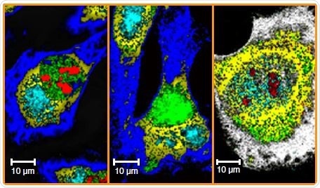
Raman microscopy for cellular investigations — From single cell imaging to  drug carrier uptake visualization - ScienceDirect
Raman microscopy for cellular investigations — From single cell imaging to drug carrier uptake visualization - ScienceDirect
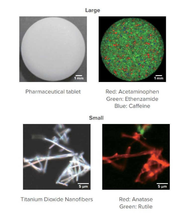
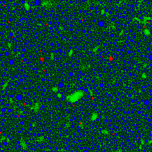
Confocal Raman Imaging in Pharmaceutical Research

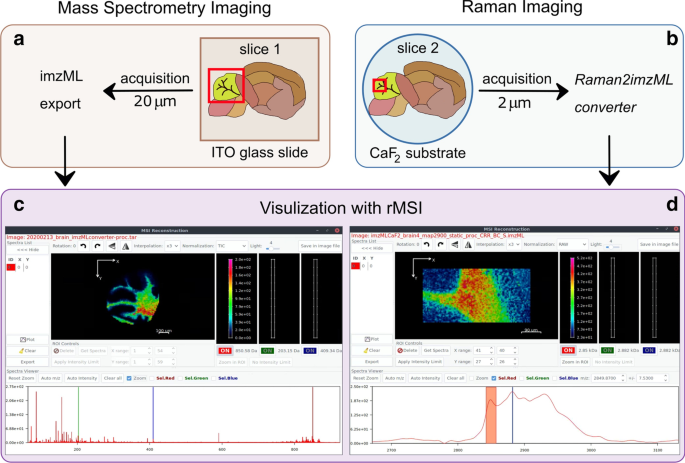
Raman2imzML converts Raman imaging data into the standard mass spectrometry  imaging format | BMC Bioinformatics | Full Text
Raman2imzML converts Raman imaging data into the standard mass spectrometry imaging format | BMC Bioinformatics | Full Text
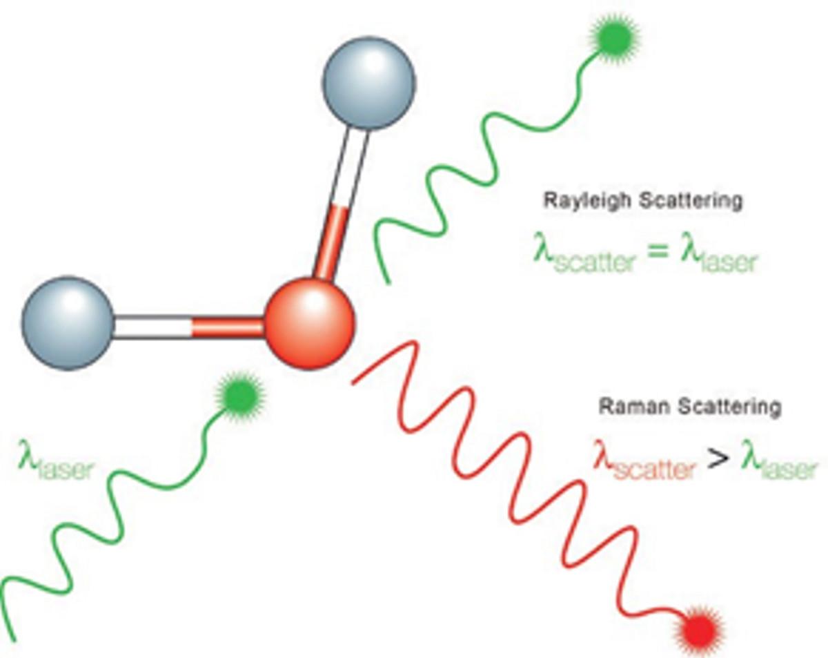
What is Raman Spectroscopy? - HORIBA

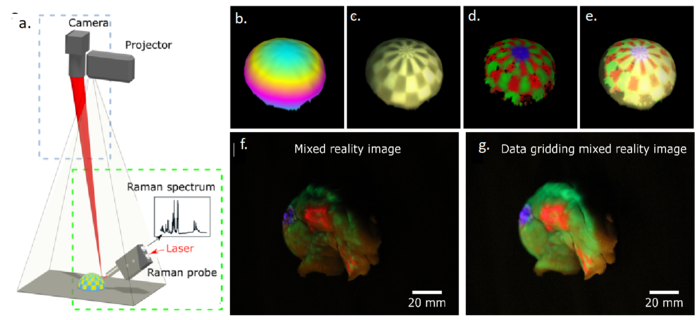
Real-time molecular imaging of near-surface tissue using Raman spectroscopy  - 2022 - Wiley Analytical Science
Real-time molecular imaging of near-surface tissue using Raman spectroscopy - 2022 - Wiley Analytical Science
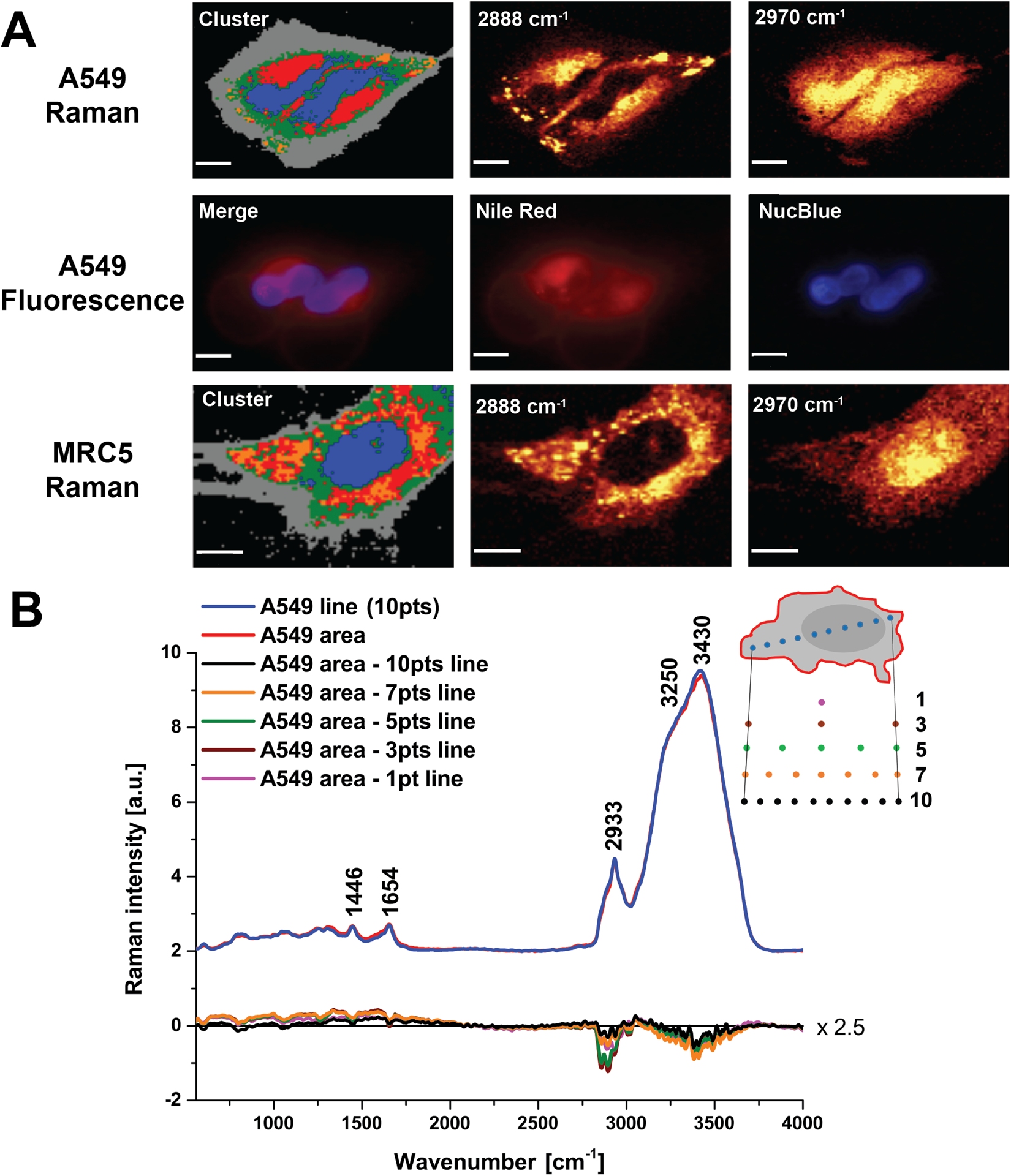
Raman Imaging - an overview | ScienceDirect Topics

Ultra-Fast Raman Mapping on Graphene - Live Demo - YouTube

High-Throughput Chemical Imaging May See Raman Microscopy in the Clinic |  Technology Networks
High-Throughput Chemical Imaging May See Raman Microscopy in the Clinic | Technology Networks

Raman images explained

Principles of Raman spectroscopy (2) Features of Raman spectoscopy | JASCO  Global
Principles of Raman spectroscopy (2) Features of Raman spectoscopy | JASCO Global

Raman Imaging Techniques | CARS, SERS, SWNTs

MCR results from Raman imaging on benign and tumourigenic uterine... |  Download Scientific Diagram
MCR results from Raman imaging on benign and tumourigenic uterine... | Download Scientific Diagram

Raman imaging of the G peak intensity (a), 2D peak intensity (b),... |  Download Scientific Diagram
Raman imaging of the G peak intensity (a), 2D peak intensity (b),... | Download Scientific Diagram

Raman Imaging Opens View to Diagnosis and Discovery | Features | Sep/Oct  2020 | BioPhotonics
Raman Imaging Opens View to Diagnosis and Discovery | Features | Sep/Oct 2020 | BioPhotonics

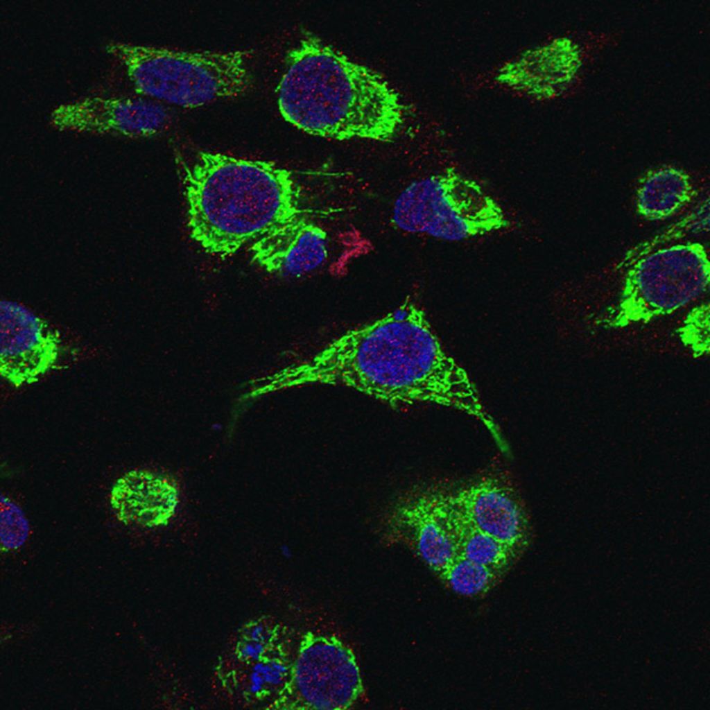
Raman micro-spectroscopy for accurate identification of primary human  bronchial epithelial cells | Scientific Reports
Raman micro-spectroscopy for accurate identification of primary human bronchial epithelial cells | Scientific Reports

Raman Imaging as a Tool for Characterizing Carbon Nanomaterials | American  Laboratory
Raman Imaging as a Tool for Characterizing Carbon Nanomaterials | American Laboratory

Raman imaging of native and W-SA sample (WPG = 40%) showing the... |  Download Scientific Diagram
Raman imaging of native and W-SA sample (WPG = 40%) showing the... | Download Scientific Diagram

Stain Free Imaging of Chemical Distribution in Biological Systems

Real time Raman imaging to understand dissolution performance of amorphous  solid dispersions - ScienceDirect
Real time Raman imaging to understand dissolution performance of amorphous solid dispersions - ScienceDirect

Raman Spectroscopy | TOPTICA Photonics AG

Applications of Raman, CARS and SRS imaging in dosage form development -  European Pharmaceutical Review
Applications of Raman, CARS and SRS imaging in dosage form development - European Pharmaceutical Review

Raman microscope - Wikipedia

Guía de Imagen Raman | Bruker

Raman spectroscopy and imaging of hASCs. The average Raman spectra of... |  Download Scientific Diagram
Raman spectroscopy and imaging of hASCs. The average Raman spectra of... | Download Scientific Diagram
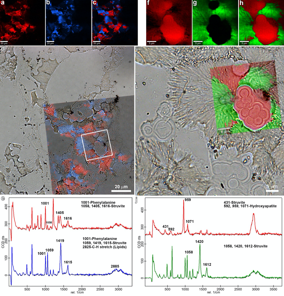
Confocal Raman Spectroscopy, Atomic Force Microscope and Scanning Nearfield  Optical Microscope- from WITec Alpha 300 Series | Carl R. Woese Institute  for Genomic Biology
Confocal Raman Spectroscopy, Atomic Force Microscope and Scanning Nearfield Optical Microscope- from WITec Alpha 300 Series | Carl R. Woese Institute for Genomic Biology
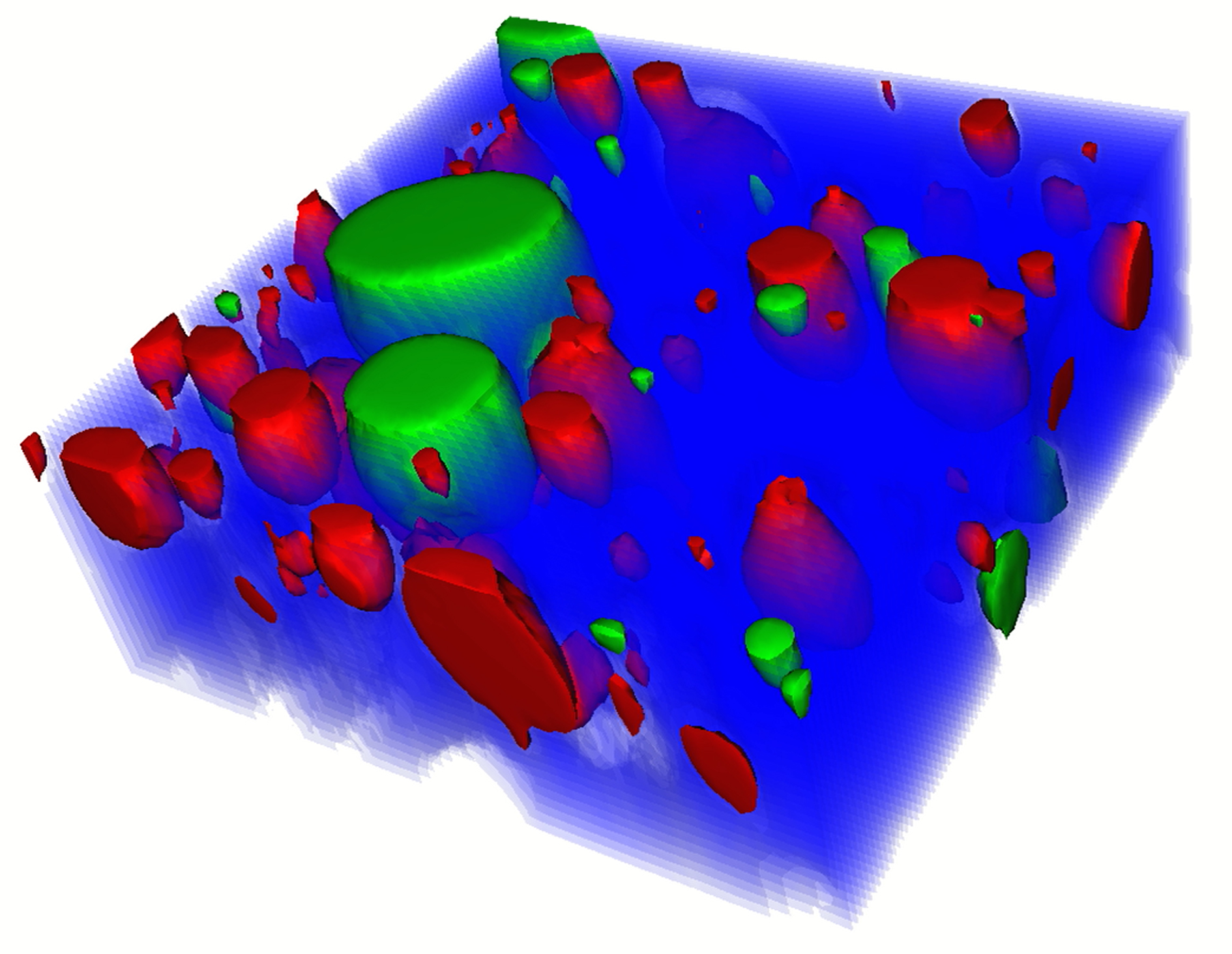
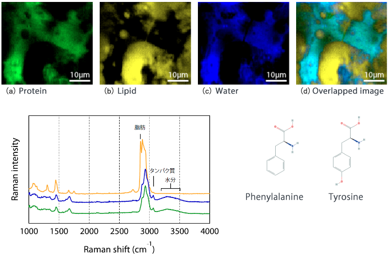
Component distribution observation of cheese by Raman spectroscopy |  Nanophoton
Component distribution observation of cheese by Raman spectroscopy | Nanophoton

PDF] The potential of Raman microscopy and Raman imaging in plant research  | Semantic Scholar
PDF] The potential of Raman microscopy and Raman imaging in plant research | Semantic Scholar

Raman spectroscopy for biomolecular analysis and label-free Raman imaging -  2020 - Wiley Analytical Science
Raman spectroscopy for biomolecular analysis and label-free Raman imaging - 2020 - Wiley Analytical Science